method
Clinical visual intelligence

Where surgical expertise becomes visual intelligence.

Method learns from the work, methodology, and outcomes of leading surgeons to support surgical education and patient visualization.

The Method
01Cases

Historical before-and-after work enters the system.

Documented cases from a surgeon’s own practice, standardised frontal, oblique and lateral views at defined follow-up intervals, enter Method under written consent. A face identifies a person, so a case is held as identifiable data throughout. Selection is recorded with the same structure: what made this patient a candidate, and what was weighed against it. Nicotine use, anticoagulants, uncontrolled hypertension, a low-lying hyoid, thin photoaged skin, and expectations no vector will meet each alter the plan, defer it, or end it. The operation that was not offered is part of a surgeon’s method.

02Intent

The surgeon’s markings, target regions, and clinical reasoning define what is intended to change.

Intent is captured as structure: the direction of lift, the plane of dissection, the regions addressed and the reasoning behind them. In this case: lengthen the neck and jawline by deepening the cervicomental angle; refresh the jawline, mouth and eyes; restore balance.

03Methodology

Patterns across the surgeon’s cases begin to form a surgeon-specific intelligence layer.

Across a documented series the same decisions recur: where tissue is released, along which vector it is repositioned, how much change is sought and where anatomy is left alone. That pattern belongs to one surgeon.

04Education

That intelligence can teach: the method becomes a structured course of cases.

Surgeons learning from an expert move between an outcome and the reasoning that produced it, case by case, annotation by annotation, without the friction of a lecture hall or a single visit to the operating room.

05Visualization

It can preview: a patient explores how this surgeon might approach someone like them.

A case-informed visualization, bounded by the surgeon’s documented series and by defined clinical constraints, with its provenance attached: which surgeon, how many cases informed it. Never a promise.

06Planning

It can support the surgeon’s own planning: a new case read against their documented series.

Comparable historical cases, the vectors and regions chosen in them, and the outcomes that followed, available while the next case is being planned, in the surgeon’s own terms.

07Capture

Toward richer capture: depth scans join the photographic record.

Millimetre-level surface geometry of the face, captured with structured-light or LiDAR depth sensors alongside the standard views, lets intent and outcome be measured, not only seen. Angles, distances and volumes become part of the case, in the surgeon’s own terms.

08Reconstruction

From points to surface: a dense scan becomes a measurable model.

Tens of thousands of surface points are rastered into a continuous mesh at sub-millimetre pitch. Holes are closed, noise is filtered, and the surface is checked against the photographs taken in the same session, so what is measured is the face, not the sensor.

09Analysis

Surface analysis: where the geometry changed, and by how much.

Curvature and deviation maps show where a surgeon’s intent met the anatomy: the jowl flattened, the submental angle deepened, the malar region left alone. Each region carries a measured difference in millimetres rather than an impression. Geometry is read alongside validated outcome: FACE-Q from the patient, a global aesthetic improvement scale from the assessor.

10Registration

Registered follow-up: the same face, aligned across time.

Every capture is registered to its baseline on stable fiducials: tragus, nasion, menton, gonion. Pre-operative, plan and follow-up then sit in one coordinate frame. Change is then a difference between aligned surfaces, comparable across cases and across surgeons. Follow-up does not stop at a year, and regions do not hold equally: in the published literature the jowl keeps most of its correction at five and a half years, while much of the gain in the cervicomental angle is given back.

Frankfort horizontalN–MeRegion · lower face and neckCervicomentalLift vectorNasolabialG–SnSn–MeDepth scannearfarsurface pointsRasterJowlSubmentalMalarflathigh · Δ mmRegisteredFollow-up50 mmGlabellaNasionPronasaleSubnasaleLabrale superiusPogonionMentonTragusGonion
Model 01 · Sculptural form · Oblique 45°
Sculptural form · oblique 45°Solid observed · dashed planned
Schematic form · posed to the documented view · not a patient
Model 02 · Surface-registered anatomy · profile 90°
Frankfort horizontalMasseterZygomaticus majorPlatysmaSMASOrbicularis oculi123456Danger zones · facial nerve1 Temporal br. · Pitanguy’s line2 Zygomatic br. · anterior parotid border3 Buccal br. · deep to the SMAS4 Marginal mandibular br. · at the border, deep to platysma5 Great auricular n. · McKinney’s point6 Cervical br. · deep to platysma below the angleDashed · Parotid outline · fixed SMASLateral canthusZygomatic archMastoidZygomatic lig.Masseteric lig.Mandibular lig.Platysma-auricular lig.Submental if neededABC
Skin · surface formSurface-registered · SMAS · muscle fibre · ligament fans · nerve branches 1–5 · A B C planes of entry · arrows lift vectors · circles release · ear path incision
11Surgeon intelligence

Every surgeon has a method.

Different surgeons may approach the same anatomy, procedure, or aesthetic goal differently: in the plane they work in, the ligaments they release, the direction they move tissue, how they treat the neck, where they place the incision. Method is designed to preserve and interpret those differences through each surgeon’s own historical cases and methodology.

Beneath the skin

A facelift is not a skin operation. Beneath the skin and its subcutaneous fat lies the SMAS, the fibromuscular sheet continuous with the platysma in the neck. It invests the mimetic muscles. Deeper lie the masseter under the parotidomasseteric fascia, and the ligaments that anchor the face to the skeleton.

SMASSuperficial musculoaponeurotic system. The layer a lift repositions
MusclesWithin the SMAS: zygomaticus major and minor · orbicularis oculi and oris · depressor anguli oris · platysma. Deeper, a muscle of mastication: masseter
LigamentsZygomatic · masseteric · mandibular · cervical. Released or left intact, by approach
Fibre direction

Each muscle runs from an origin to an insertion, and the tissue over it descends, and is repositioned, in relation to that line.

MasseterA muscle of mastication, not a mimetic muscle. Zygomatic arch and maxilla → angle and ramus of the mandible. The superficial head runs down and back, the deep head near vertical. The SMAS glides over the parotidomasseteric fascia that covers it.
ZygomaticusZygomatic body → modiolus at the corner of the mouth. The line the nasolabial fold deepens against.
PlatysmaFascia over the clavicle and upper chest → mandible and lower face; fibres run up and forward. Its laxity and medial banding shape the ageing neck.
OrbicularisConcentric fibres around the orbit and the mouth. Oculi has an orbital part over the rim and a palpebral part in the lids. Sphincters, not lifters.
Retaining ligaments

Ligaments tether the soft tissue to bone or to deep fascia. The facial nerve branches run deep to them, so release stays superficial to the nerve. Which are released, and which are kept, is one of the defining choices of an approach.

ZygomaticOsteocutaneous · McGregor’s patch, anterior-inferior to the arch · released to mobilise the midface
MassetericFasciocutaneous · anterior border of the masseter, upper and lower · released in the extended deep plane
MandibularOsteocutaneous · parasymphyseal, about 1 cm above the border · released to smooth the jowl; preserved in some techniques
Platysma-auricularFasciocutaneous · from the parotid fascia to the skin in front of the ear · defines the fixed SMAS
Danger zones

The plane and depth of dissection are chosen around the facial nerve. Method records where an approach works in relation to it.

TemporalPitanguy’s line, 0.5 cm below the tragus to 1.5 cm above the lateral brow; superficial as it crosses the zygomatic arch
Zygomatic · buccalEmerge at the anterior border of the parotid, deep to the SMAS. The branches a sub-SMAS dissection works above
Marginal mandibularAt or above the inferior border of the mandible behind the facial vessels, below it in front. Deep to the platysma throughout
Great auricularMcKinney’s point, 6.5 cm below the ear canal on the sternocleidomastoid. The nerve most often injured in facelift
Approach A · Extended deep plane

One interpretation of the same anatomy: release everything that tethers the composite, then move it vertically.

PlaneSkin raised subcutaneously in front of the ear, then a sub-SMAS composite flap entered along the line from the angle of the mandible toward the lateral canthus, passing over zygomaticus major
VectorVertical · ≈75° to the Frankfort horizontal
ReleaseZygomatic · masseteric · mandibular · cervical retaining ligaments
NeckPlatysma addressed in continuity with the SMAS; lateral suspension to the mastoid
SubmentalOpened when the midline limits the result: subplatysmal fat, anterior platysma, and the digastric and submandibular gland where they define the floor
IncisionRetrotragal
Approach B · High SMAS

A second: enter higher, above the zygomatic arch, and carry the flap superolaterally.

PlaneBilamellar: the skin raised off the SMAS as one flap, then the SMAS raised as a second flap entered above the zygomatic arch, angling posteriorly at the anterior ear. The two are moved on different vectors
VectorSuperolateral · ≈55° to the Frankfort horizontal
ReleaseZygomatic · masseteric ligaments
NeckLateral platysma suspension to the mastoid
SubmentalAdded only where lateral suspension cannot reach the midline
IncisionPretragal
Approach C · SMAS plication

A third: no deep dissection. The SMAS is folded and sutured, and change is concentrated where the fold is.

PlaneSkin elevated; SMAS folded on itself and sutured at the fixed-mobile junction, no sub-SMAS dissection. Imbrication and lateral SMASectomy are the neighbouring options
VectorOblique · ≈35° to the Frankfort horizontal
ReleaseNone. Retaining ligaments left intact
NeckMidline platysmaplasty via a submental approach
SubmentalCentral to this approach: corset platysmaplasty after subcutaneous and subplatysmal lipectomy. Whether the medial bands decussate decides how far the midline must be opened
IncisionRetrotragal

The same procedure does not mean the same approach.

Method preserves the differences between surgeons: their methodology, visual preferences, and outcome patterns. It does not average them away. The three approaches shown are illustrative technique families from the published literature, drawn schematically. Lateral SMASectomy, the MACS lift and SMAS imbrication are omitted here. They are not attributed to any participating surgeon and are not clinical advice.

12Volumetric anatomy

The same anatomy, in depth.

Model 03 builds each structure beneath the skin as a solid: the SMAS as a sheet, the muscles as bellies with thickness, the facial nerve as a course that runs under the SMAS and emerges from the parotid, the ligaments as bundles from bone to dermis. A dissection window opens on the lateral face so the layers can be read in order, and compared with the surface-registered model above.

Dissection window

Skin and subcutaneous fat are lifted over the lateral face. The first layer beneath is the SMAS: fixed over the parotid, mobile in front of it.

SkinAbout 3–5 mm with the subcutis · lifted within the window
FatSuperficial compartments over the SMAS: medial, middle and lateral temporal cheek, nasolabial, jowl. The fat a lift repositions
SMASFibro-fatty sheet at roughly 5 mm · continuous with the platysma below and the temporoparietal fascia above
ParotidSuperficial lobe beneath the fixed SMAS · the facial nerve divides inside it
Muscles and bone

Within the SMAS: the mimetic muscles, built as bellies with thickness. Deeper: the masseter, a muscle of mastication under its own fascia, and the two bones a facelift works against.

MasseterMastication, not mimetic · zygomatic arch and maxilla → angle and ramus · about 1 cm thick · the SMAS glides over the parotidomasseteric fascia
ZygomaticusZygomatic body → modiolus · major and minor · the line the nasolabial fold deepens against
Depressor a. o.Mandible → modiolus · draws the commissure down
PlatysmaClavicular fascia → mandible and lower face · lateral and medial bands · shown to the base of this scan’s short neck
OrbicularisOculi and oris · sphincters around the orbit and the mouth
FasciaParotidomasseteric fascia over the masseter: the floor of the sub-SMAS plane, with the nerve branches beneath it
DeepTemporalis beneath the deep temporal fascia, which splits above the arch into two layers around the superficial temporal fat pad · sternocleidomastoid · buccinator and the buccal fat pad
Neck at depthDeep to the platysma: subplatysmal fat, the anterior belly of digastric, and the submandibular gland. These decide whether a neck can be improved and how far
BoneZygomatic arch and mandible: origin, insertion, and the border the marginal nerve follows
Fibre direction

Striations run along each muscle from origin to insertion. Descent and repositioning are read against them.

MasseterDown and back
ZygomaticusDown and forward, to the corner of the mouth
PlatysmaUp and forward, from the clavicle to the mandible
OrbicularisConcentric
Retaining ligaments

Bundles from bone or deep fascia to the dermis, shown at depth. Osteocutaneous ligaments are denser; fasciocutaneous ones sparser.

ZygomaticOsteocutaneous · the zygomatic cutaneous ligaments along the zygomatic body, with McGregor’s patch behind them · released to mobilise the midface
OrbicularisOrbital rim periosteum to skin through orbicularis oculi · marks the lid-cheek junction · short and sparse
MassetericFasciocutaneous · anterior border of the masseter, upper and lower
MandibularOsteocutaneous · parasymphyseal · released to smooth the jowl, kept in some techniques
Platysma-auricularFasciocutaneous · parotid fascia to the skin in front of the ear · the fixed SMAS
Facial nerve

The trunk enters the parotid, divides at the pes anserinus, and its branches leave the gland deep to the SMAS, becoming more superficial as they run forward.

1 TemporalWithin the temporoparietal fascia as it crosses the arch · Pitanguy’s line
2 ZygomaticAnterior parotid border → orbicularis oculi · deep to the SMAS
3 BuccalOver the masseter, beneath the parotidomasseteric fascia → mouth
4 Marginal mandibularAt or above the border behind the facial vessels, below it in front · deep to the platysma
5 Great auricularOn the sternocleidomastoid, directly beneath the SMAS-platysma · McKinney’s point
6 CervicalDeep to the platysma below the angle of the mandible · the branch a facelift most reliably meets
Depth, compared

What the volumetric model adds over the surface-registered one above.

OcclusionStructures hide and reveal one another correctly as the head turns
PlanesSubcutaneous, supra-SMAS and sub-SMAS planes are separable: the plane an approach enters can be shown, not implied
DepthNerve branches run beneath the SMAS and emerge from the parotid at clinical depth
MeasurementThickness, distance and depth become measurable rather than painted
LimitsBuilt on a placeholder head from its landmarks · not patient-specific · for review by a surgeon before use
SMAS · fibro-fatty sheetParotid · superficial lobeLateral temporal-cheek fatMiddle cheek fatMedial cheek fatSubplatysmal fat · midlineJowl fatMasseterZygomaticus majorDepressor anguli orisPlatysma · lateral bandPlatysma · medial bandOrbicularis oculiOrbicularis orisTemporalis · under the deep temporal fasciaDeep temporal fascia · splits above the archParotidomasseteric fascia · floor of the sub-SMAS planeDigastric · anterior belly · subplatysmalSubmandibular gland · subplatysmalSternocleidomastoidBuccal fat pad · buccinator beneathZygomatic archMandibleMcGregor’s patch · zygomatic ligaments arise within itZygomatic cutaneous ligs. · osteocutaneousOrbicularis retaining lig. · lid-cheek junctionMasseteric lig. · upperMasseteric lig. · lowerMandibular lig. · osteocutaneousPlatysma-auricular lig. · fasciocutaneous · fixed SMASFacial nerve · trunk · pes anserinus1 Temporal br. · Pitanguy’s line2 Zygomatic br.3 Buccal br.4 Marginal mandibular br.6 Cervical br. · deep to platysma5 Great auricular n. · McKinney’s point
Model 03 · Volumetric anatomy · dissection window
Skin · surface form · obliqueIvory fascia and bone · oxblood muscle · ochre nerve · copper ligament
Placeholder head · anatomy placed from its landmarks at clinical depth · schematic, for surgeon review
13Selected cases

Documented the same way, case by case.

Each participating surgeon’s work enters Method through the same structure: the pre-operative view, the plan drawn on that view, and the outcome at follow-up. Photography is shown as clinical documentation, with consent. Never as advertising.

Case series 01 · Face and neck

A deep-plane face and neck lift, read against its plan.

The plan is drawn on the pre-operative view itself, so the outcome can be read against what was intended: where tissue was released, along which vector it was repositioned, and where the neck was redefined.

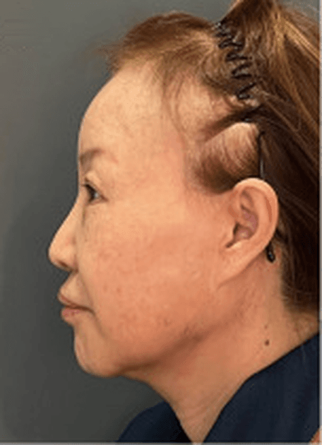
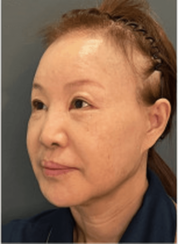
Extensive work was done to lengthen the neck and jawline by increasing the definition in the internal angle of the neck, more than she had even in her youth. The jawline, mouth and eyes look more refreshed, and the face has a better balance after receiving the lifts.
Operating surgeon’s note
Procedure
Deep-plane face and neck lift
View
Left oblique 45°
States
Before · Intent · Outcome
Pre-operative photograph, oblique viewPost-operative photograph, oblique view
Region · lower face and neckTragusCervicomental angleLift vector
Fig. 01 · Before · oblique 45°
Drag to move between states
14Documentation

Five views, one standard.

Method’s documentation standard follows established clinical photography conventions, so that cases from different practices can be compared on equal terms, and so that a change in the photograph is a change in the patient, not in the camera.

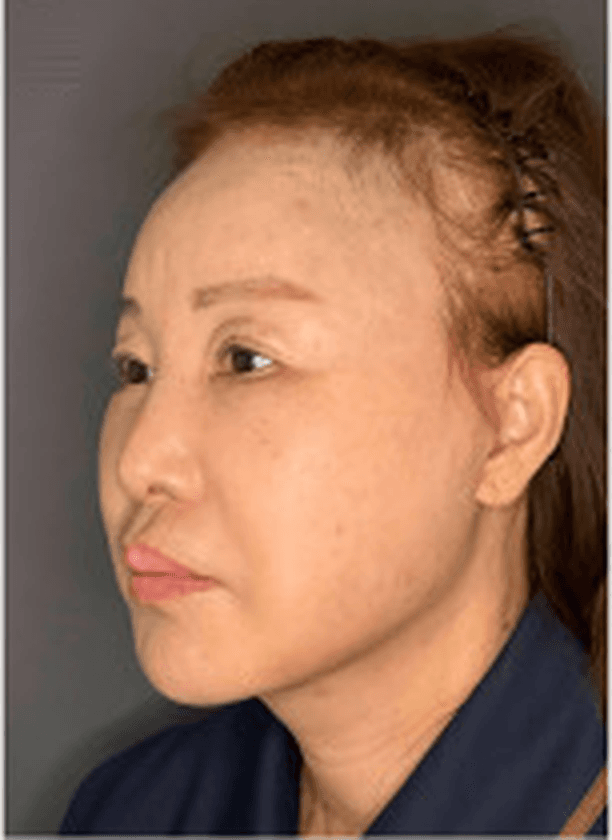
Frontal pre-operative photograph, case series 01
Fig. 03 · Frontal · 0°Case 01
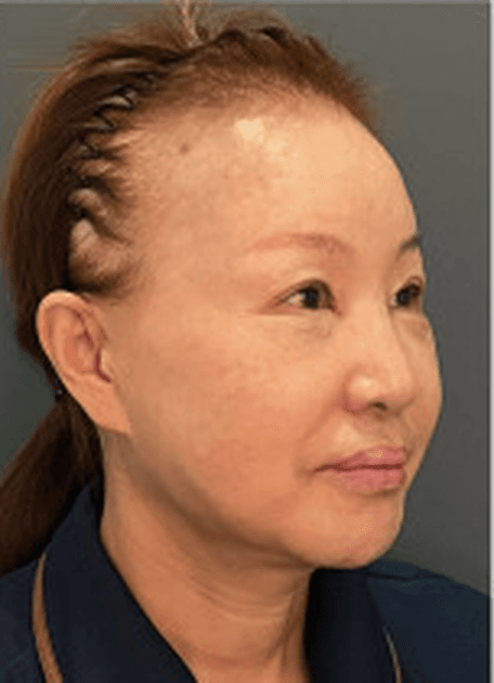
Right oblique pre-operative photograph, case series 01
Fig. 04 · Right oblique · 45°Case 01
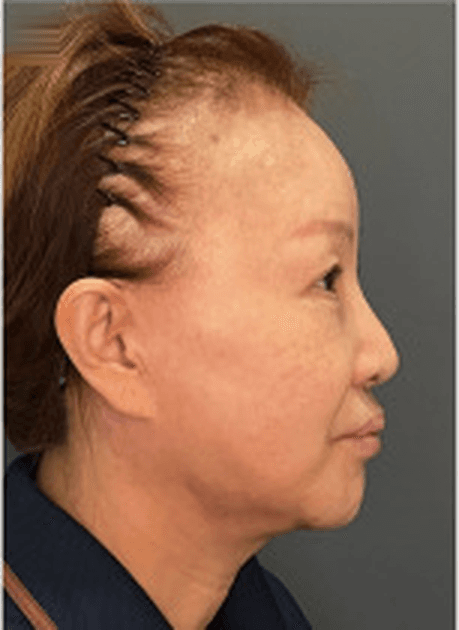
Right lateral pre-operative photograph, case series 01
Fig. 05 · Right lateral · 90°Case 01
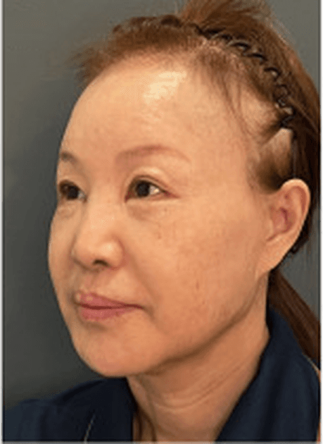
Left oblique pre-operative photograph, case series 01
Fig. 06 · Left oblique · 45°Case 01
Left lateral pre-operative photograph, case series 01
Fig. 07 · Left lateral · 90°Case 01
01
Head position

The Frankfort horizontal, from the top of the tragus to the infraorbital rim, is held level, with a neutral expression. Neck extension is avoided: on its own it can mimic a lift.

02
Capture

A prime lens of 90 to 105 mm on full frame, at a fixed subject distance of about 1.5 m. Shorter focal lengths bring the camera closer for the same framing and enlarge whatever is nearest the lens. Two diffused sources at 45 degrees to the subject, 5000 to 5500 K, at fixed positions. The same five views every time.

03
Backdrop

Plain, non-reflective and mid-tone, with the subject set well clear of it so no shadow falls across the outline.

04
Preparation

Hair off the face and ears. Make-up, jewellery and glasses removed. Clothing kept clear of the region documented, and the same at every session: a collar over the cervicomental angle breaks the comparison.

05
Animation

The five standard views are recorded in repose. Function is recorded as well: a smiling view, and a view of the neck with the platysma contracted. Platysmal banding is often visible only on contraction.

06
Intervals

Pre-operative, then follow-up at three, six and twelve months. Nothing earlier than three months is read as an outcome: residual oedema and an immature scar still shape the photograph. Only like-for-like views at like-for-like intervals are compared.

07
Alteration

Photographs are not retouched, filtered, smoothed or relit. Crop, framing and aspect ratio are held constant between the pre-operative view and follow-up: recropping on its own can read as a change in the jawline.

08
Adjuncts

Anything else done in the same session is recorded with the case: blepharoplasty, brow lift, fat grafting, resurfacing, filler. An outcome is attributed to the lift only when the record shows the lift was done alone.

09
Course

The post-operative course is recorded with the case: haematoma and any return to theatre, sensory or motor nerve disturbance and its recovery, skin ischaemia, scar quality, and alopecia at the temporal incision. Haematoma is the commonest early complication of a face lift.

10
Consent

Every case enters Method under written consent, given separately for each use: the clinical record, teaching, publication, and use in a case-informed preview. A full-face photograph identifies the patient, so a case is held as identifiable data throughout and is never described as anonymised. Direct identifiers are removed. Consent can be withdrawn, and the case with it.

15Two applications

One intelligence layer. Two applications.

Everything below is powered by the same surgeon-specific layer. It is one platform: what a surgeon teaches and what a patient previews come from the same documented method. The diagram below traces that single source into its two uses.

Documented series · plan · outcome
Surgeon intelligence layer
→ Teaching the method
Previewing the method →
For surgeons
In development with participating surgeons

Preserve, structure, and communicate methodology.

Method helps a surgeon’s reasoning outlive the lecture, case by case, annotation by annotation.

  • 01TeachingProcedure collections that move from indication to plan to outcome.
  • 02Case-based learningRead a result against the decision that produced it.
  • 03AnnotationsMarkings, regions, vectors and measurements kept with the case.
  • 04Visual reasoningWhy this approach, for this anatomy, in the surgeon’s own terms.
  • 05Knowledge transferA method that can be examined after the operating room closes.
For patients
Planned

Explore a surgeon’s method, applied to you.

Patients can explore visualizations informed by the historical work and methodology of a participating surgeon.

  • 01Surgeon-specific visualizationHow might this surgeon approach someone like me?
  • 02Case-informed previewInterpreted through documented cases, within clinical constraints.
  • 03Comparable historical casesReal, consented cases with anatomy similar to yours.
  • 04Educational explanationWhat is being changed, what is not, and why.

A visualization is a case-informed preview, a surgeon-specific visual interpretation. It is not a prediction, a guaranteed outcome, or medical advice, and it does not replace a consultation.

16Clinical intelligence

Not image generation.

The technical challenge is not to produce an image. It is to produce a reliable one: an output that stays inside what is clinically defined and can be traced back to the surgeon it came from.

Every output must
  • 01Reflect surgeon-specific patterns
  • 02Remain within defined clinical constraints
  • 03Preserve anatomy that should not change
  • 04Avoid uncontrolled model drift
  • 05Remain interpretable
Method does not
  • Generate from a prompt, or from a generic model of appearance
  • Alter anatomy outside the surgeon’s documented scope
  • Present any output as a predicted or guaranteed result
  • Represent a surgeon’s method without that surgeon’s review
01
Input

Consented, standardised clinical photographs and the surgeon’s plan for the case.

02
Constraint

Defined clinical bounds. Anatomy that should not change is constrained; change is limited to the regions the surgeon addresses.

03
Surgeon intelligence

Patterns learned from one surgeon’s documented series: vectors, regions, degree of change.

04
Visualization

An interpretable, surgeon-specific preview that carries its provenance: which surgeon, how many documented cases informed it.

17Governance

Approached clinically, built carefully.

Method handles clinical photographs of real people. The rules below are not a policy page; they are constraints the system is built around.

01
Consent first

Cases enter Method only with documented patient consent for the stated purpose. Consent can be withdrawn, and the case with it.

02
Identifiable by nature

A face identifies a person. Masking the eyes does not change that. Every case is held as identifiable data under documented consent, never as anonymous. Names, dates and locations are never displayed.

03
Surgeon review

Each participating surgeon reviews how their methodology is represented before anything is shown to others. A clinician sees any patient-facing preview before the patient does.

04
Clinical constraints

Change is bounded by the regions and degrees documented in the surgeon’s series. Nothing outside them is generated.

05
Provenance

Every preview names the surgeon whose method informed it and the size of the documented series behind it.

06
Narrow before wide

One procedure family, a small group of practices, and evaluation with clinicians before anything expands.

07
Health data

A clinical photograph is data concerning health: special category data under GDPR Article 9, processed only under explicit consent, stored encrypted with access limited to the treating team and named reviewers.

08
Retention

Every case has a defined retention period agreed at consent. At its end, or on withdrawal, the images are deleted and the case is excluded from any future training.

09
Synthetic images

Every case-informed preview is labelled as generated, not photographed, at the point it is shown. It is never presented beside a photograph without that label.

10
Adults only

Method is not shown to, and does not process cases from, anyone under 18.

11
Regulatory status

The proof of concept is an educational and documentation tool. Planning support that would influence a clinical decision is a medical-device function, and will be assessed as one before it is offered.

18Vocabulary

The terms Method is built on.

Method describes surgery in the surgeon’s own language. These are the terms that appear in plans, annotations and previews, defined once and used consistently.

SMAS
The superficial musculoaponeurotic system: the fibromuscular layer beneath the skin of the face, continuous with the platysma in the neck. The layer a deep-plane lift repositions.
Deep plane
The dissection plane beneath the SMAS. Working here releases the retaining ligaments so skin and SMAS move together as one composite, without tension on the skin.
Retaining ligaments
Fibrous anchors from bone or deep fascia to the skin. The zygomatic and mandibular ligaments are osteocutaneous; the masseteric and platysma-auricular ligaments arise from deep fascia. Their release determines how far tissue can be repositioned.
Lift vector
The direction along which released tissue is repositioned: vertical, oblique or posterior. One of the clearest signatures of a surgeon’s method.
Platysma
The paired thin sheet of muscle running from the fascia over the clavicle up to the mandible and lower face, continuous with the SMAS. Its medial edges are the bands that show with age. Its laxity and banding shape how the neck ages.
Cervicomental angle
The angle between the underside of the chin and the front of the neck. The published criterion for a youthful neck is 105 to 120 degrees. Its definition is a primary index of neck rejuvenation.
Gonial angle
The angle at the gonion between the posterior border of the ramus and the inferior border of the mandible. Its soft-tissue definition sharpens the jawline.
Jowl
Descended tissue gathering along the jawline as the supporting layer loosens with age.
Tragus
The small cartilage in front of the ear canal. The posterior reference of the Frankfort horizontal, and the anchor toward which lift vectors are drawn in Method’s plans.
Frankfort horizontal
The horizontal reference plane from porion to orbitale, taken on the skin as the line from the tragus to the infraorbital rim, held level for every standardised photograph.
Documented series
A surgeon’s consented cases, captured to one standard, with plan and follow-up. The unit Method learns from.
Case-informed preview
Method’s term for a visualization: a generated image, not a photograph, interpreting one surgeon’s documented series applied to a new face. Educational, not predictive.
Facial ageing
Four processes that a lift addresses unevenly. Descent of soft tissue as the retaining ligaments attenuate. Deflation as the fat compartments lose volume. Skin change: thinning, loss of elasticity, photodamage. Bone resorption at the orbit, the maxilla and the mandible. A lift repositions and tightens; it does not restore volume or skin quality.
Five layers
The frame every structure sits in, from the surface down: skin, subcutaneous fat, the SMAS, the retaining ligaments and the spaces between them, and the deep fascia over bone and the muscles of mastication. The facial nerve runs deep to the fourth layer in the lateral face and becomes more superficial as it runs forward.
Fixed and mobile SMAS
Behind the platysma-auricular ligament, over the parotid, the SMAS is fixed. In front of it, over the masseter and the cheek, it is mobile. The junction is where every SMAS technique does its work.
Facial nerve
The seventh cranial nerve. Its trunk enters the parotid, divides at the pes anserinus, and its five branches (temporal, zygomatic, buccal, marginal mandibular, cervical) leave the gland deep to the parotidomasseteric fascia and the platysma.
Danger zone
A region where a nerve runs superficially enough to be injured by dissection: the temporal branch on Pitanguy’s line as it crosses the arch, the great auricular nerve at McKinney’s point on the sternocleidomastoid, the marginal mandibular branch at the mandibular border, the cervical branch below the angle.
Nasolabial angle
The angle at subnasale between a tangent to the columella and a tangent to the upper lip. Published norms are about 90 to 95 degrees in men and 95 to 110 in women.
Facial thirds
Conventionally trichion to glabella, glabella to subnasale, and subnasale to menton, each about a third. The G–Sn and Sn–Me readouts on the form report the middle and lower thirds as a share of the glabella-to-menton height.
N–Me
Anterior facial height: the distance from nasion to menton, the standard vertical dimension in cephalometry.
Landmarks
Glabella: the most prominent point of the forehead between the brows. Nasion: the deepest point of the nasal bridge at the root. Pronasale: the tip of the nose. Subnasale: where the columella meets the upper lip. Labrale superius: the midpoint of the upper vermilion border. Pogonion: the most anterior point of the chin. Menton: the lowest point of the chin. Gonion: the angle of the mandible. Tragion: the notch above the tragus.
Plication, imbrication, SMASectomy
Three ways of tightening the SMAS without a sub-SMAS flap. Plication folds it on itself and sutures the fold. Imbrication overlaps a cut edge and sutures it. Lateral SMASectomy excises a strip at the fixed-mobile junction and closes the gap. The MACS lift suspends the SMAS with purse-string sutures from the deep temporal fascia.
19Questions

Asked often, answered plainly.

No. It is an educational preview informed by a specific surgeon’s documented cases: a visual interpretation of how that surgeon has approached similar anatomy. It is not a guarantee, a simulation of your surgery, or medical advice.

Participating surgeons, clinical collaborators, advisors and partners. The surgeon education tools are in development with participating practices; patient-facing tools are planned and will be introduced with the same constraints.

The proof of concept begins with one procedure family, the face and neck, where methodology differs clearly between surgeons and outcomes are well documented. Coverage grows as documented series are contributed.

By contributing a consented, standardised case series of photographs, plans and follow-up, and reviewing how their methodology is represented before it is shown to anyone else.

The contributing surgeon and their patients. Use is limited to the purposes agreed at contribution, and a case can be withdrawn: its images are removed and it is excluded from any future training.

No. Method does not generate from prompts or from a generic model of appearance. Every output is bounded by one surgeon’s documented series and by defined clinical constraints, and carries its provenance.

A face and neck lift is surgery. Haematoma is the commonest early complication. Others include temporary or, rarely, lasting weakness from injury to a branch of the facial nerve, numbness from the great auricular nerve, skin ischaemia at the flap edge, widened or visible scars, hair loss at the temporal incision, asymmetry, and the need for revision. Outcome also varies with healing and with the anatomy each patient brings. A preview does not change any of this, and it is not a substitute for a consultation.

20Direction

Where this can go.

Stated as direction, not commitment. The proof of concept comes first.

  • 01Surgeon-specific models
  • 02Richer 3D capture
  • 03Scanning
  • 04Structured teaching systems
  • 05Case intelligence
  • 06Visual surgical planning
  • 07Patient education

Every great surgeon has a method.
Method makes it visible.

Method is in early development with participating surgeons, clinical collaborators and advisors. If you are a surgeon with a documented series, a clinician, a researcher or a partner who would like to be introduced to the project, request access.